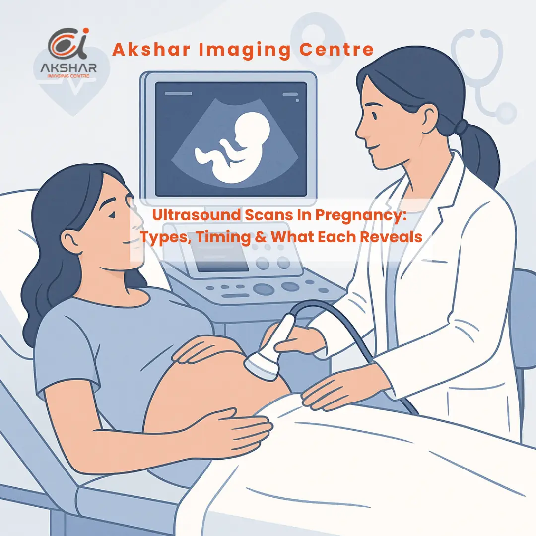
Pregnancy ultrasound scans utilize sound waves to generate a picture of your baby on a screen. A certified medical professional, pregnancy care provider, or doctor will use it to check your baby’s health.
It also helps you determine if you are at risk of pregnancy complications. Parents can also track the baby’s growth. It can capture snapshots of their movements and monitor the baby’s heartbeat.
In the journey of pregnancy, these not only give you a sigh of relief knowing your baby is healthy but also build a deep personal connection through these very first interactions.
However, it is essential to know what to expect from ultrasound scan pregnancy tests, know about the types such as transvaginal, abdominal, or Doppler ultrasound, and much more. And that’s what we explore in this article!
What Are Ultrasound Scans In Pregnancy?
An ultrasound scan in pregnancy, or a sonogram, is a prenatal diagnostic test during pregnancy that examines the health and development of your baby.
An obstetrician or an ultrasound technician conducts these tests for various reasons. Many times, ultrasounds are performed to check on your baby and make sure they’re growing properly. Other times, the doctors or pregnancy care provider will ask for an ultrasound after they detect a problem.
How Ultrasound Scans In Pregnancy Work?
When an ultrasound is performed, the sound waves are sent through the patient’s vagina or abdomen using a device called a transducer. The sound waves hit the structure inside the body, including the baby and the reproductive organs.
These sound waves are then converted into images in the machine, which the sonographer can display on the screen. There is no use of radiation, such as X-rays, to visualize the baby in this case.
What Can an Ultrasound Scan Pregnancy Detect?
Here are the reasons why your prenatal ultrasound scan is performed:
- Whether to confirm you’re pregnant or not
- Determine the gestational age and the due date of the unborn baby
- Determine whether you have a molar pregnancy, ectopic pregnancy, miscarriage, or other complications of pregnancy
- Find out whether it is multiple babies, i.e, twins, triplets, or more
- You can check the baby’s position in the uterus
- See the location of the placenta
- Check the baby’s growth, heart rate, and movement
- Examine pelvic organs like the ovaries, uterus, and cervix
What Are The Types Of Ultrasound In Pregnancy?
1. Transvaginal (Endovaginal) Ultrasound
A transvaginal ultrasound is a variation of the regular pelvic ultrasound scan, whereby, in the process, your pregnancy healthcare professional will have to insert an object into your vagina. It is similar to how you place a tampon. It helps to confirm the pregnancy, and also:
- Determine gestational age
- Identify ectopic pregnancy
- Locate placenta
- Assess the potential miscarriage risk
This scan test is usually conducted during the first trimester, which is between 1st and 12 weeks, and can last 15 minutes to an hour.
2. Abdominal Ultrasound
Abdominal ultrasound can also be referred to as Trans abdominal ultrasound. In this instance, the provider of medical care places a transducer on your skin. They then rotate this equipment around your belly to capture images of your baby.
They will often apply a bit of pressure to get the best view. This type of ultrasound scan in pregnancy is suggested after 12 weeks of gestation, especially for morphology or anatomy scans.
3. Doppler Ultrasound
Your physician could prescribe a Doppler ultrasound scan, used to detect blood flow to the baby. In most cases, it will take place between 2 and -40 weeks. It helps get more information on the blood flow in fetal organs, placenta, and umbilical cord.
4. 3D / 4D Ultrasound
3D and 4D ultrasound offer volumetric, lifelike images useful for assessing facial structures or suspected anomalies. Often adjunctive; not a routine for all pregnancies.
5. Anatomy Scan
An anatomy scan, also known as an anomaly scan, is a stage II or 20-week scan that examines the fetal anatomy and growth. It monitors the growth of the unborn organs and other body structures, as well as identifying some kinds of congenital disabilities.
6. Nuchal Translucency (NT) Scan
This scan is carried out between 11 and 14 weeks, at the end of the first phase, also known as the first trimester. This scan takes a reading of the fluid around the fetal neck to screen against the possibility of chromosomal disorders such as Down, Edward, and Patau syndromes.
Timing & Standard Schedule of Ultrasounds And What Each Scan Reveals
Early Pregnancy: 5 to 8 weeks
A gestational sac can be detected through transvaginal ultrasound within 5 weeks, a yolk sac within 5.5 weeks, and a beating embryo within 67 weeks.
6 weeks to 8 Weeks
Abdominal ultrasound may detect a heartbeat if it is more than or equal to 8 weeks, but for earlier detection, you should prefer transvaginal imaging.
11 weeks to 14 Weeks
An NT scan is performed for chromosomal screening and dating.
18 weeks to 22 Weeks
Anatomy/morphology scan with thorough examination of fetal structure, location of the placenta, levels of fluids, and sex (if requested).
Third Trimester
Should there be any complications, i.e., in the case of a placenta problem, hindrance in growth, or anything of the sort, then your healthcare provider will advise further scans after you enter into the third trimester of the pregnancy, or an ultrasound scan during your 9th month.
According to standard recommendations, at least two scans throughout your pregnancy are expected.
Conclusion
Ultrasound scans are routinely required during pregnancy to ensure fetal growth and health, and to examine for potential complications. It is a non-invasive way through which medical professionals can monitor the health and growth of your baby.
You can also see the baby’s movements through these scans, figure out whether you’re pregnant with one or multiple babies, estimate due date, and so much more to make this journey better.
We at Akshar Imaging ensure every scan is performed with precision, care, and the latest technology, supporting healthy beginnings for both mother and baby.
FAQ’s
Q. How Many Ultrasound Scans Should You Expect In Pregnancy?
A. Generally, in case you have a healthy, regular, and uncomplicated pregnancy, you will have approximately 3 to 4 ultrasound tests. In case any problems come up, your doctor can prescribe further scans.
Q. How Can You Read a Ultrasound Scan Pregnancy Report?
A. Be sure that you master the universal abbreviations of such reports, which are as follows: GA, EDD, LMP, et cetera. You must search for the gestational age heartbeat, growth sizes of the placenta, position, and fluid levels. Nevertheless, you should never forget to consult your doctor.
Q. How Early Can An Ultrasound Detect Pregnancy?
A. A transvaginal scan can show a sac at 5 weeks and a heartbeat by 6–7 weeks. Abdominal scans are detected later.
Q. When Is a Doppler Ultrasound Scan Performed In Pregnancy?
A. The most common time for a Doppler ultrasound during pregnancy is the second trimester, as it is vital for evaluating fetal growth and well-being.
Q. Can an Ultrasound Scan Detect Pregnancy?
A. Yes. It verifies a pregnancy inside the uterus and displays the heartbeat, typically beginning from 5−7 weeks of the period.

