At Akshar Imaging Centre, we provide advanced cervical X-ray services to help diagnose neck, spine, and upper back conditions with precision and care.
A cervical X-ray is a simple diagnostic imaging test that produces clear images of the cervical spine (the neck region of your backbone). It allows doctors to evaluate the bones, alignment, and surrounding structures of the neck. This test is commonly used to detect injuries, degenerative changes, and other conditions affecting the cervical spine.
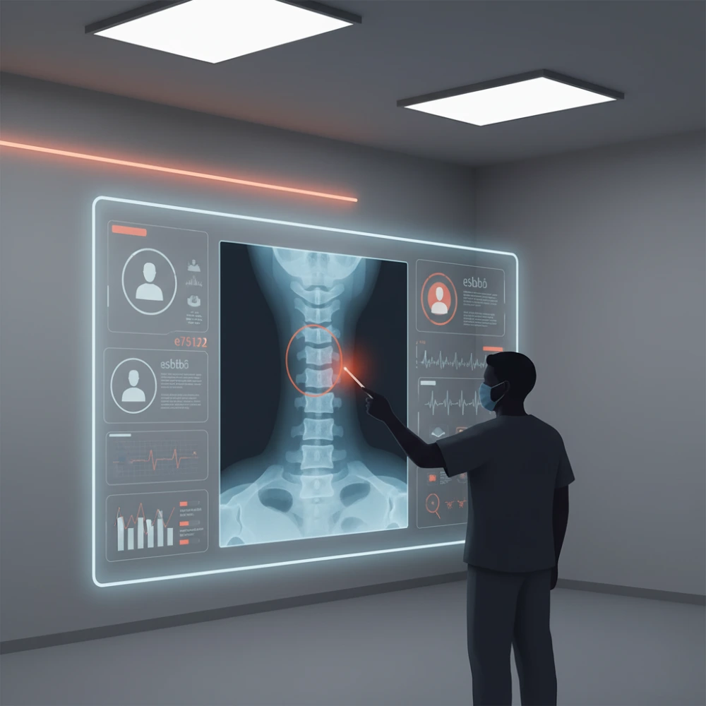
A cervical X-ray uses low-dose radiation to capture images of the vertebrae in your neck. It shows the bones, spinal alignment, and sometimes soft tissues around the cervical spine. It is quick, painless, and an important tool in diagnosing neck pain, stiffness, or trauma.
A radiologist examines the X-rays for fractures, dislocations, bone spurs, degenerative changes, vertebrae alignment, arthritis, infections, or abnormal growths. Results are quickly prepared and shared with your doctor for diagnosis and treatment planning.
Benefits: Quick, non-invasive, detects fractures, spine disorders, and provides a baseline for treatment. Low radiation exposure compared to CT scans.
Risks: Small radiation dose, limited detail for soft tissues compared to MRI/CT, not suitable during pregnancy unless essential.
Yes, it involves a very small amount of radiation. We take extra precautions for children and avoid it during pregnancy unless essential.
The procedure usually takes around 5–10 minutes.
It may be to check for fractures, arthritis, abnormal curvature, or causes of persistent neck pain.
It clearly shows bones and alignment issues. For soft tissue or nerve-related problems, your doctor may suggest MRI or CT.
Founded by Dr.Riddhesh Panchal, the centre has offered accurate & advanced imagining services continuously for last 15 years.
Ground Floor Shop No.1 & 6, Unique Metropolis, Nr. R.C. Technical Road, Opp. Prasang Party plot, Gota, Ahmedabad-382481
+91 7069 777277, 7069 777377
aksharic@gmail.com